What Is A Periapical XRay and Why Do You Take It?
posted: Oct. 27, 2022.
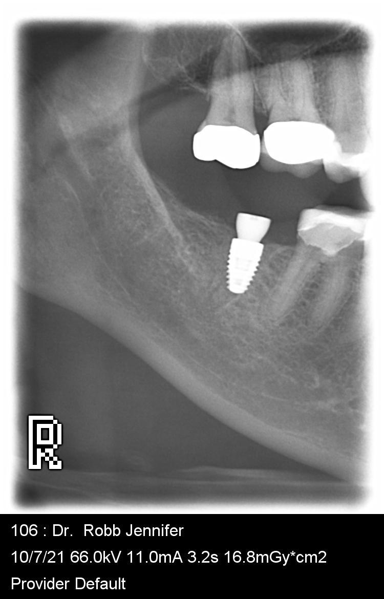
A periapical xray is an xray that shows your tooth from the chewing surface to the end of the root of the tooth and the bone surrounding that tooth. Dentists use periapical xrays to check for abscesses (usually seen as a dark area around the end of the tooth root), to check the bone levels around your tooth, and to check for dental decay. Sometimes small areas of decay will not show on this type of xray. The focus of this xray is on the root of the tooth, so sometimes the chewing surfaces of the teeth overlap slightly and hide small areas of decay.
Periapical Xrays can be taken with a sensor or film placed in your mouth or they can be taken with a larger machine that allows your dentist to block out areas that she or he does not need to see while still taking the xray of the tooth or teeth that are in the area of concern. The larger xray machine often only has a bite stick that you bite on with your front teeth and very little is in your mouth. (The picture above is one that was taken with a panoramic xray machine where all but the area for these 2 to 3 teeth was blocked out.